New Webinar from Prof. Cáceres – SOLVED

Prof. Cáceres is preparing a new webinar about the “Pulmonary fat, my friend”. Join him this Wednesday June 16th at 12:00 CEST. He will discuss the case presented here, among others. Register here.

Dear Friends,

Today’s radiographs belong to two different patients with peripheral opacities of the chest. What would be your diagnosis?

1. A is fat and B solid tissue
2. B is fat and A solid tissue
3. Both are fatty
4. Need a CT

Click here to see the answer

Findings: Radiograph of case A shows an opacity in the left upper lung. Its inner contour is outlined by air (arrow) and the outer border is not visible (asterisk) suggesting an extrapulmonary lesion (incomplete border sign).
CT shows that the opacity represents extrapleural fat (B, arrow).

Radiograph of the second case shows a well-defined rounded opacity (C, arrow), that was interpreted as a peripheral pulmonary nodule. PET-CT was done, and the apparent pulmonary nodule was shown to be extrapleural fat (D, arrow).

 
These two cases are shown to emphasize that fat in or around the lung cannot be distinguished from soft tissues in the plain chest radiograph. To recognize fat, CT is necessary.
 
More information about fatty lesions of the lung is given in the webinar “Mediastinal fat: my friend” that will be published soon on the EBR youtube channel.

Dr. Pepe’s Diploma Casebook 171 – SOLVED

Dear Friends,

presenting a new case of “Big little findings”. Radiographs belong to a 62-year-old man diagnosed of colon carcinoma one year ago. Talc pleurodesis performed after discovering right pleural implants.

What do you see? 

Click here to see the answer

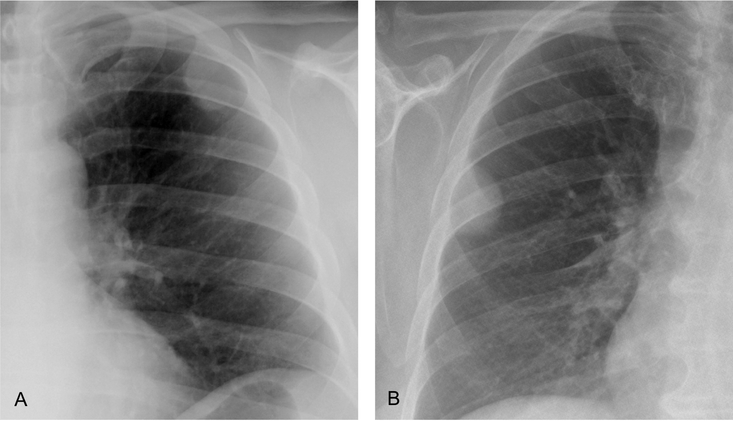
Findings: PA radiograph (A) shows a right pleural effusion, secondary to talc pleurodesis. The lateral view shows D9 loss of height with erosion of the inferior vertebral plate (B, circle). The findings are partially obscured by the superimposed pleural effusion and are better seen in the insert (C).

Comparison with a sagittal CT taken six months earlier confirms that the chest radiograph findings were not present at that time (D and E, circles).

Coronal and sagittal CT show crumbling of D9 (A and B, circles). There is air in the intervertebral space, which goes against infection. MRI confirms the findings (C and D, circles). Final opinion was metastasis vs. compression fracture. Given the lack of trauma and the presence of metastases in other organs, metastasis was considered the best diagnosis. No further action was taken.

Final diagnosis: metastasis to D9 (unproven)

I am presenting this case to emphasize, once again, the importance of looking at the thoracic spine, an important landmark in the chest radiograph. Hidden by the mediastinal structures in the PA view, it is clearly depicted in the lateral radiograph.
It is important to check the spine in each lateral view because it can offer information that may be overlooked.

This case includes three basic points to remember when reading chest radiographs:

1. Satisfaction of search. The pleural effusion centers our attention and prevents examining other areas that may show important findings.
2. Comparison with previous films. Very useful to demonstrate that the finding is real and was not present previously.
3. Performing a thorough checklist. Discovering the abnormal vertebra takes a conscious effort of analysis of the lateral view, a routine that should not be forgotten.

Once the spinal abnormality is found, cross-sectional imaging (CT and/or MRI) is the method of choice to confirm the findings and reach a likely diagnosis.

To reinforce this concept, I am showing three more cases of spinal disease that might have been missed if we had not paid attention to subtle findings.

CASE 1. 73-year-old woman with back pain for one month. Lateral chest shows a compression fracture of D12 (A, circle), partially hidden by the diaphragm. The fractured vertebra is better seen in the cone-down view (B). Compression fractures of vertebral bodies are related to osteoporosis and common in advanced age. They cause significant pain, leading to inability to perform daily activities. If they are not recognized, they lead to a decline in the well-being of elderly patients.

CASE 2. 57-year-old man with back pain. Initial film shows D9 height loss that was overlooked (A, arrow). Three months later there is obvious collapse of D9 (B, arrow). CT confirms the collapsed vertebra and irregularity of the D10 upper plate (C, circle). Diagnosis: tuberculosis

CASE 3. 34-year-old man with back pain and fever. PA chest film (not shown) was uninformative.
Lateral view shows increased opacity of the middle third of the thoracic spine and an indistinct D7-D8 space (A, circle). Findings are more evident in the cone-down view (B).

Sagittal CT shows irregularity of the intervertebral disk and erosion of the end plates (C, circle).
Coronal and axial CTs show soft-tissue involvement, responsible for the increased opacity in the lateral chest film (D and E, arrows).
Diagnosis: infectious spondylitis


Follow Dr. Pepe’s advice:

Remember to look at the thoracic spine in the lateral radiograph. You may see subtle findings that portend relevant disease.

This is the way the world ends
Not with a bang but a whimper
(T S Eliot)

Dear friends,
This is our last case. For diverse circumstances Dr. Pepe and I have decided to abandon the EBR blog. We hope you’ve enjoyed the cases and that they’ve contributed to your education. Thanks for the interest you have shown over the years.
Our best wishes to you all.

Cáceres’ Corner Case 256 – SOLVED

Dear Friends,

welcome to the second trimester of 2021! Showing today PA chest radiograph of a 66-year-old man with chest pain without any other symptoms.

What do you see?
More images will be shown on Tuesday and Wednesday.

Dear friends, showing today the lateral chest view.
Does it help?

Today I am showing an enhanced axial CT.
What would be your diagnosis?

Click here to see the answer

Findings: PA chest radiograph shows an increase in size and opacity of the left hilum
(A, arrow), due to superimposition of a well-defined posterior mass visible in the lateral view (B, arrow). At first glance, the appearance of the mass is compatible with an extrapulmonary lesion. However, there is retrocardiac nodule in the PA view (A, red arrow), suggesting a metastasis from an intrapulmonary mass.

Enhanced axial CT confirms an irregular pulmonary mass (C, arrow), which is invading the chest wall, as confirmed by the displaced intercostal artery (C, yellow arrow) and erosion of the underlying rib (D, circle).

Caudal slices confirm the retrocardiac nodule (E, white arrow) and additional nodules (E-F, red arrows) representing pleural implants.
Biopsy of the main mass returned as lung carcinoma.

Final diagnosis: Carcinoma of the lung simulating an enlarged hilum in the PA view.
 
Congratulations to Dr LeLam and thaf1212, who detected the retrocardiac nodule, which is the clue to determine that the main mass is intrapulmonary.
 
Teaching point: Remember that one of the three causes of unilateral enlarged hilum is superposition of a pulmonary opacity either in front or behind the hilum (the other two are enlarged hilar lymph nodes and increase in size of the pulmonary artery)

Dr. Pepe’s Diploma Casebook 170 – SOLVED

Dear Friends,

This week’s case follows the pattern of a “Meet the Examiner” presentation, with questions and answers similar to a real examination. Take your time before scrolling down for the answer.

There will be no new blog posts over the Easter period. The next case will be published on Monday, April 5, 2021.

The images belong to a 65-year-old woman with cough and low-grade fever. The referred physician demanded a chest CT.

What would be your diagnosis?

1. Pneumonia
2. Pulmonary infarction
3. Peripheral adenocarcinoma
4. Any of the above

Click here to see the answer

Findings: unenhanced axial and sagittal CTs show LLL airspace disease with a surrounding halo (B-C arrows). In my opinion, the sensible answer is 4. Any of the above, although I liked adenocarcinoma because of the peripheral halo and air bubbles within the infiltrate (A, circle).

Click here to see more images

Patient was diagnosed of pneumonia and treated with antibiotics, without improvement. Chest radiographs taken 13 days later shows progression of the LLL opacity (A and B, arrows).

A CT was recommended.

Click here to see the CT images

Two axial and one sagittal views are selected. What would your diagnosis be:

1. Peripheral adenocarcinoma
2. Tuberculosis
3. Covid pneumonia
4. None of the above

Click here to see the answer

In comparison with the previous CT, the LLL infiltrate has increased markedly in less than two weeks. An upper halo persists (A and C, arrows). A small infiltrate has appeared at the right lung base (B, arrow) In my opinion, this rapid progression rules out carcinoma and TB. A PCR was negative. Blood tests were not remarkable. It was considered that the patient had an unusual pneumonia, and the antibiotic was changed.

Click here to see more images

The fever disappeared with the new antibiotic and the patient improved moderately. A new CT was taken three weeks later. What would your diagnosis be?

1. Löffler syndrome
2. Goodpasture syndrome
3. Cryptogenic organizing pneumonia
4. Any of the above

Click here to see the answer

Findings: The most striking finding is the disappearance of the LLL infiltrate and the apparition of two new areas of airspace disease in RLL and LLL (A, arrows). There is a halo sign in the LUL infiltrate (B, arrows) and a negative halo in the RLL infiltrate (B and C, arrows).
This change of location of the opacities falls in the category of migratory infiltrates which are caused by several diseases, some of them listed in the previous questions.

The patient had no risk factors for parasitic infection and no peripheral eosinophilia, ruling out Löffler syndrome. Renal function was not altered, excluding Goodpasture’s syndrome

The combination of migratory infiltrates and a negative halo sign was very suggestive of a cryptogenetic organizing pneumonia, that was confirmed with BAL and an excellent response to corticosteroid treatment.

Final diagnosis: cryptogenic organizing pneumonia

Organizing pneumonia (OP) is a clinical, radiological and histological entity usually associated to other pathologies. The idiopathic form of OP is called cryptogenic organizing pneumonia (COP).
Clinical manifestations of COP begin with a mild flu-like illness with fever, cough and malaise.
In chest imaging it may appear as localized airspace opacity that may be confused with ordinary pneumonia, adenocarcinoma or aspiration, among others. The lack of response to antibiotic treatment and the peripheral location may help in suggesting the diagnosis.

I am presenting this case because it shows two features the help in the diagnosis: migratory infiltrates and the reverse halo sign.
Migratory infiltrates are not unique to COP, but they occur in a limited number of diseases (Loeffler syndrome, vasculitis, etc.) and their presence in the adequate clinical setting should suggest COP.
The reverse halo was originally described as specific of COP, but since then it has been seen in many other entities. It is defined as a central ground-glass opacity  surrounded by denser consolidation of crescentic shape or a complete ring. It is visible in about 20% of cases.

In this patient the combination of both signs strongly pointed towards COP, that was confirmed and responded brilliantly to corticoid treatment.

To complete the presentation, I am showing two more examples of reversed halo and migratory infiltrates (CASES 1 and 2, below).

CASE 1. 61-year-old woman with COP and basilar infiltrates (A, arrows). During treatment, coronal and axial CTs show bilateral and symmetrical reversed halo signs (B and C, arrows)

CASE 2. 51-year-old woman with COP and migratory pulmonary infiltrates (A and B). The second CT shows nice examples of reversed halo sign (B, circle), better seen in the cone down axial view (C, arrows).


Follow Dr. Pepe’s advice:

1. Localized cryptogenic organizing pneumonia may mimic other pulmonary processes

2. Migrating infiltrates and reverse halo sign (or both) are helpful in suspecting COP

Cáceres’ Corner Case 255

Dear friends,

today I am presenting preoperative chest radiographs for knee surgery in a 47-year-old woman.

More images will be shown on Wednesday.

What do you see?

Click here to see the new images

Click here to see the answer

Findings: PA chest radiograph shows a bump in the left hemidiaphragm (A, arrow). It is partially hidden in the lateral view by the shadow of the right hemidiaphragm and the cardiac silhouette (B, arrows).

Diaphragmatic bumps are common on the right and rarer on the left, especially in young persons. I was curious about this finding and reviewed an abdominal CT done a few weeks earlier. Enhanced axial, coronal and sagittal images demonstrate an intact diaphragm and a fluid-filled structure in the thoracic side (C-E, arrows). The appearance is typical of a diaphragmatic cyst.
 
Diaphragmatic cyst is a congenital lesion, asymptomatic and absolutely harmless. It is easy to demonstrate with CT and should not be removed. They are rare (I have seen only four during my professional life). I thought it interesting to acquaint you with this rare entity.

Final diagnosis: congenital diaphragmatic cyst
 
Teaching point: not all diaphragmatic bumps are hernias or eventrations. When they occur in the left side in a young person, consider other possibilities, such as a congenital cyst or a fibrous pleural tumor.

Cáceres’ Corner Case 254 – SOLVED

Dear Friends,

today’s radiographs belong to a 34-year-old woman with moderate cough. Previous history of asthma.

What do you see?

Diagnosis:

1. Mucous plug
2. Segmental atelectasis
3. Tuberculosis
4. None of the above

Click here to see the answer

Findings: Pa chest radiograph shows a tubular opacity that seems to arise from the right hilum (A, arrow). The lateral chest (B) does not show any abnormality, which raises the possibility that the opacity in the PA view is spurious.

Careful inspection demonstrates that the opacity extends to the right apex and to the neck (C, red arrows). The appearance is typical of a superimposed pigtail.

Some of you described the slightly elevated minor fissure. It is an unfortunate coincidence, probably related to previous episodes of mucous plug in an asthmatic patient causing mild loss of volume of RUL.
 
Final diagnosis: Pigtail simulating pulmonary disease.
 
Congratulations to MK who was the only one to suggest the correct diagnosis.
 
Teaching point: You may think that I tricked you, but it was not my intention. This case is a reminder that apparent pulmonary opacities may be located in the pleura, chest wall or outside of the body.
 
To emphasize this point I am showing two more cases of braids simulating pulmonary disease, presented in earlier blogs.

CASE 1. 48-year-old woman with mild cough. PA radiograph shows an ill-defined opacity in the left lung, running from top to bottom (A, white arrows). The opacity extends towards the neck (A, red arrow), which suggests that it is external to the lung. Lateral view shows an elongated opacity in the back of the chest (B, arrows).

A photo of the patient (C) confirms that a long braid is the cause of the opacity. PA radiograph after lifting the braid demonstrates that the chest is normal (D).

CASE 2. 25-year-old man with braided hair simulating a RUL infiltrate (A, arrow). The opacity extends to the neck, giving away the diagnosis (A, red arrow). After raising the braid, the chest looks normal (B). Remember that men also wear their hair long nowadays.

Dr. Pepe’s Diploma Casebook 169 – SOLVED

Dear Friends,

Presenting a new case of “Big little findings”. Preoperative chest radiograph for meniscus surgery in a 56-year-old woman.

What do you see?

Click here to see the answer

Findings: PA view shows a small right hemithorax. There is elevation of the right hemidiaphragm and a small hilum (A, red arrow). The findings are very suggestive of RLL lobectomy. The oblique fissure in the RLL represents the displaced minor fissure (A, white arrow). Previous CT shows a normal-size right lung with a ground-glass opacity in the RLL (B, arrow).

Final diagnosis: RLL lobectomy for adenocarcinoma of the lung

I am showing this case to discuss displacement of the lung fissures, an important finding that can indicate partial collapse of the underlying lobe. Usually, lobar collapse is detected because of the increased opacity of the lobe. Occasionally, the collapsed lobe retains much of its air, so a shift of the fissure may be the only sign of collapse.

A potential pitfall of fissure displacement is previous surgery, as seen in the case presented. In my experience, excluding previous surgery, aerated lobar collapse occurs mainly in the following conditions:

1. Inflammatory peripheral lung disease
2. Central lobar bronchial obstruction
3. Rounded atelectasis

NORMAL ANATOMY
The right minor fissure is visible in about 50% of chest radiographs as a straight horizontal line at the level of the right hilum (Fig. 1, A and B) The right and left major fissures are not visible in the PA film because their course is not tangential to the x-ray beam. (A, curved dotted lines). They are both visible as oblique lines in the lateral view (B).

Fig. 1
Fig. 2. PA radiograph showing the minor fissure (A, arrow). The lateral view shows both the right minor and major fissures (B, white arrows) and the upper portion of the left major fissure (B, red arrow)

Inflammatory lesions can cause scarring which diminishes the size of the affected lobe. TB is the most common cause in upper lobes. Bronchiectasis is the predominant cause in lower lobes. Both conditions can show an aerated lobe with loss of volume (Figs. 3-5).

Fig. 3. 68-year-old woman with previous history of TB. There is aerated partial collapse of RUL as evidenced by the elevated minor fissure (A and B, white arrows). Fibrotic changes are seen in the apex (A and B, red arrows). An incidental finding is calcification of breast prostheses.
Fig. 4. RLL collapse secondary to bronchiectasis. There is an oblique line at the right base (A, white arrow) that simulates an inferior accessory fissure. However, the right hilum is markedly low (A, red arrow), indicating loss of volume of RLL. Coronal CT shows marked RLL collapse with bronchiectasis, outlined by the displaced major fissure (B, arrow).
Fig. 5. 56-year-old man with previous TB. Lateral view shows forward displacement of the left major fissure (A, arrows), indicating partial collapse of LUL. PA radiograph depicts marked elevation of left hilum (B, arrow), secondary to fibrotic TB.

Central lobar bronchial obstruction is occasionally associated with aerated lobar collapse. It is thought to be due to collateral air ventilation through incomplete fissures (Figs. 6-7).

Fig. 6. Routine follow-up of an 82 y.o. man who underwent surgery for laryngeal carcinoma 10 years ago. PA view shows abnormal left hilum and blurring of the left cardiac contour (A, arrow). Lateral view shows marked forward displacement of the left major fissure (B, arrows) indicating severe LUL collapse.

Unenhanced axial CT confirms the marked LUL collapse (C, white arrow) secondary to endobronchial obstruction (C, red arrow). CT taken one year earlier shows an endobronchial lesion (D, red arrow) and discrete forward displacement of the major fissure (D,E, white arrows). These changes were overlooked. Surgical diagnosis: bronchogenic carcinoma

Fig. 8. Aerated RLL collapse in central carcinoma. PA radiographs shows a very low right major fissure (A, red arrow), better depicted in the cone down view (B, arrow). The left hilum is descended (A, white arrow). Bronchoscopy discovered a carcinoma of the RLL bronchus. The oblique line in the RUL corresponds to a scar.

Rounded atelectasis is a common cause of fissure displacement. It occurs secondary to spiral folding of the lung parenchyma when fixed by thickened pleura. The consequence is a peripheral rounded opacity in an aerated collapsed lobe. The volume loss, detected by the displaced fissure, avoids possible confusion with a true nodule in the plain film.

Fig. 9. Asymptomatic 49-year-old man with rounded atelectasis. Notice the visibility of the left major fissure, indicating LLL volume loss (A, arrows). Lateral view shows an ill-defined posterior opacity which corresponds to the rounded atelectasis (B, arrow).

Axial and sagittal CT confirm displacement of the left major fissure (C and D, white arrows), the small LLL, and the posterior rounded atelectasis (C, red arrow).

As a final thought, occasionally you may find fissure displacement without an apparent cause (Fig. 10).

Fig. 10. 92-year-old man, asymptomatic. PA radiograph shows downward displacement of the minor fissure (A, white arrow), major fissure (A, yellow arrow) and right hilum (A, red arrow). In a previous film four years earlier, the minor fissure (B, yellow arrow) and the right hilum (B, red arrow) were moderately descended. Since the patient was 92 y.o. and had no symptoms, his physician decided not to do a CT scan. My impression is that he has fibrotic changes in the RLL, which is not unusual in advanced age.


Follow Dr. Pepe’s advice:
1. A displaced fissure may be the only manifestation of aerated lobar collapse (always exclude previous surgery).

2. Most common causes:

a) Peripheral lobar inflammatory disease

b) Central bronchial obstruction

c) Rounded atelectasis

Cáceres’ Corner Case 253 – SOLVED

Dear Friends,

Today’s case is a PA chest radiograph for knee surgery in a 28-year-old man.

What do you see?

Click here to see the answer

Findings: PA chest radiograph shows an osteochondroma in the right humerus (A, yellow arrow). There are two more in the anterior arch of the left fifth rib and in the proximal end of the right clavicle (A, red arrows).
They are better seen in the cone down views (B-D, arrows).

The first and only diagnosis that comes to mind is multiple osteochondromatosis, confirmed with views of the lower extremities (E-G).

Final diagnosis: Multiple osteochondromatosis.
 
Most of you did very well in this case. Congratulations to Mauro, who was the first and to Kaushalya and Ali who made back-to-back diagnosis in a five-minute interval.
 
Teaching point: remember to look at the bones of the chest, especially when taking an examination. It may surprise the examiner and win you a few extra points.

Dr. Pepe’s Diploma Casebook 168 – Solved!

This week’s case is a little special! Prof. Cáceres has prepared a Quiz that will to challenge your knowledge and your speed reviewing radiographs! The quiz contains 7 different cases with radiographs and you will have 60 seconds to answer each question! The three participants with highest score will receive a signed picture of Dr. Pepe 😀

Are you up for the challenge? Join the quiz here It will start on Thursday 18th, at 12:00 CET.

On Friday Prof. Cáceres will publish the explanation for each case on the blog.

CASE 1

CASE 1

71-year-old man with hemoptysis:

In which quadrant will you place the lesion?

1. Right upper quadrant
2. Right lower quadrant
3. Left upper quadrant
4. Left lower quadrant

Click here to see the answer

Findings: there is a rounded opacity behind the heart (A, arrow). CTs show a non-enhancing pulmonary mass with irregular borders attached to the pericardium (B-C, circles).
Post-operative diagnosis: mucoid carcinoma of the lung.


CASE 2

CASE 2


43-year-o.ld male with moderate cough.

Most likely diagnosis:

1. Tuberculosis
2. Enlarged left pulmonary artery
3. Carcinoma
4. Any of the above

Click here to see the answer

Findings: PA radiograph shows that the left hilum is larger and more opaque than the right one (A, arrow). The pulmonary arch is prominent (A, red arrow). The lateral view shows an enlarged left pulmonary artery (B, arrows) excluding the diagnosis of carcinoma or TB as causes of hilar enlargement. Enhanced axial CT confirms the enlarged left pulmonary artery (insert, arrow).
Diagnosis: Congenital pulmonary valve stenosis with secondary dilatation of the left pulmonary artery due to the jet effect.

CASE 3

CASE 3


60-year-old man with chest pain.

In which quadrant will you place the lesion?

1. Right upper quadrant
2. Right lower quadrant
3. Left upper quadrant
4. Left lower quadrant

Click here to see the answer

Findings: there is a well-defined rounded left apical opacity (A, arrow), better seen in the cone down view (B, arrow)

Coronal and axial enhanced CT confirm the apical mass (C-D, arrows). The patient complained of pain in the left shoulder. Needle biopsy came back as adenocarcinoma.

Final diagnosis: Pancoast tumor

CASE 4

CASE 4

65-year-old man with cough and dyspnea

Diagnosis:

1. Unilateral hyperlucent lung
2. Pneumothorax
3. Giant bulla
4. Carcinoma of the lung

Click here to see the answer

Findings: the initial impression of the PA chest is a left hyperlucent lung with diminished vascularity. A second look shows a descended left hilum (A, arrow) and a concave paraspinal line (A, red arrow) representing the major fissure. These findings are indicative of LLL collapse with compensatory expansion of LUL.

 
Enhanced axial CT confirms the marked LLL collapse (B, arrow). Coronal reconstruction shows irregular bronchial narrowing (C, red arrow) with complete occlusion of the LLL bronchus.

Diagnosis: epidermoid carcinoma with LLL collapse.

CASE 5

CASE 5

33-year-old man with pain in the right hemithorax

Where is the nodule located:

1. Lung
2. Pleura
3. Chest wall
4. Need a CT

Click here to see the answer

Findings: PA radiograph shows a rounded opacity in the lower right chest (A, circle) with incomplete border sign (medial aspect outlined by air, lateral border not visible because in contact with chest wall). The clue to its location is given by the rib erosion (A, red arrow) which places the lesion in the underside of the rib. The border of the erosion is sclerotic, indicating a slow-growing process.
CT confirms a soft-tissue mass (B, circle) and the rib erosion (B-C, red arrows).

Final diagnosis: neurofibroma in a patient with neurofibromatosis.

CASE 6

CASE 6


32-year-old man with chronic cough

In which quadrant will you place the lesion?

1. Right upper quadrant
2. Right lower quadrant
3. Left upper quadrant
4. Left tower quadrant

Click here to see the answer

Findings: PA radiograph shows increased lucency of the lower right lung with decreased vasculature (A, circle). This finding has two main causes: increased lung air or paucity of lung vessels (pulmonary embolism, arterial stenosis). In these cases, the best approach is to take an expiratory film, which will demonstrate whether or not there is air-trapping. If present, it will orient us to a bronchial obstructive process, either central or peripheral

Coronal CT (B) confirms the increased lucency and diminished vasculature of RLL and RML. Scattered bronchiectasis are seen within the lucent lung (B-C, arrows).
Axial expiratory CT (D) demonstrates marked air-trapping of RML and RLL.

The patient had a history of swallowing a peanut at the age of five years, developing RLL pneumonia at that time. Control radiographs demonstrated increased lucency of the lower right lung over the years.
 
Final diagnosis: Lobar Swyer-James/McLeod syndrome secondary to aspiration of a peanut in childhood.


CASE 7

CASE 7


17-year-old woman with moderate cough

Most likely diagnosis:

1. Benign pulmonary nodule
2. Arteriovenous malformation
3. Pleural plaque
4. Artifact

Click here to see the answer

Findings: PA radiograph shows a rounded opacity in the periphery of the left lung (A, arrow) that seems to be calcified. Cone down view shows a whorled pattern (B, arrow). A braid is visible in the left supraclavicular area (A, red arrow).

Scout view of the CT does not show the apparent lung lesion, which is not visible in the axial view of the lung (D). The technician that did the CT noticed that the patient had a long braid with a rubber band at the end.

Final diagnosis: hair braid simulating a lung nodule.

Cáceres’ Corner Case 252 – SOLVED

Dear Friends,

Since this week is my birthday, I am showing a simple case. Chest radiographs were taken in a routine study for asbestos exposure in a 42-year-old man.

Will show more images on Wednesday.

Click here to see the images shown on Monday


Dear Friends,

showing today a cone down view of the lateral chest. What does the pattern suggest?

Click here to see the new images

Click here to see the answer

Findings: PA radiograph shows punctate opacities in the upper and middle thirds of the right lung. The right heart border is indistinct (A, circle) suggesting RML disease.

The lateral view confirms RML disease (B, circle). A cone down view demonstrates thick lineal branching lines (C, circle) highly suspicious of dilated mucous-filled bronchi.
(Branching structures in chest radiograph are either vessels or mucous -filled bronchi).

Unenhanced sagittal and axial CTs show bronchiectasis of RML and lingula (D-E, circles).

Final diagnosis: RML bronchiectasis detected in the lateral view of the chest
 
Congratulations to MK, who made the diagnosis.
 
Teaching point: I presented this case because it is a nice example of bronchiectasis with mucous impaction suspected in the plain film. I posted it on Monday without having seen the CT because whoever read it told me that bronchiectasis were present.
I reviewed the CT two days ago and was surprised to discover two vital findings
that I had not been told:
 
1. The CT showed centrilobular and tree-in-bud opacities (F-G, circles), typical signs of bronchiolitis.
2. These findings plus RML and lingular bronchiectasis are a classic presentation of atypical mycobacterial infection.

So, what started as an unsuspected discovery in the plain film ended up with the serendipitous diagnosis of atypical mycobacterial infection (unproven, but likely). The attending physician has been notified and when a germ is found I would let you know