
Prof. Cáceres is preparing a new webinar about the “Pulmonary fat, my friend”. Join him this Wednesday June 16th at 12:00 CEST. He will discuss the case presented here, among others. Register here.
Dear Friends,
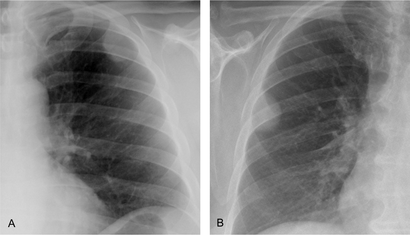
Today’s radiographs belong to two different patients with peripheral opacities of the chest. What would be your diagnosis?
1. A is fat and B solid tissue
2. B is fat and A solid tissue
3. Both are fatty
4. Need a CT
Click here to see the answer
Findings: Radiograph of case A shows an opacity in the left upper lung. Its inner contour is outlined by air (arrow) and the outer border is not visible (asterisk) suggesting an extrapulmonary lesion (incomplete border sign).
CT shows that the opacity represents extrapleural fat (B, arrow).

Radiograph of the second case shows a well-defined rounded opacity (C, arrow), that was interpreted as a peripheral pulmonary nodule. PET-CT was done, and the apparent pulmonary nodule was shown to be extrapleural fat (D, arrow).

These two cases are shown to emphasize that fat in or around the lung cannot be distinguished from soft tissues in the plain chest radiograph. To recognize fat, CT is necessary.
More information about fatty lesions of the lung is given in the webinar “Mediastinal fat: my friend” that will be published soon on the EBR youtube channel.
…..A , angolo acuto, intrapolmonare… solido .B , extrtapolmonare, grasso…US è sufficiente per integrazioine diagnostica. Grazie PROF.