


Dear Friends,
Today I am presenting chest radiographs of a 66-year-old man with cough and low-grade fever.
What would be your diagnosis?
1. Pleural effusion
2. Lobar collapse
3. Pneumonia
4. Any of the above
You have one week to post your answers. The correct answer will be given during the webinar of Tuesday 18 at 12:30 P.M.
You can join the webinar here


Click here for the answer
Findings: PA radiograph shows a descended left hilum (A, arrow). There is a triangular retrocardiac opacity (C, red arrow) and blunting of the left costophrenic angle (A, yellow arrow). These findings are typical of LLL collapse, as many of you mentioned (see Diploma case 63).
The lateral view shows a posterior triangular shadow (B, arrow) which represents the collapsed LLL.

Coronal CT confirms the triangular shaped LLL collapse (C, red arrow), causing elevation of the left hemidiaphragm (C, arrows). There is no evidence of pleural effusion.
Axial CTs show a mass at the origin of the LLL bronchus (D-E, arrows). Note the markedly collapsed LLL (D-E, red arrows).
Bronchoscopy and biopsy confirmed the diagnosis of carcinoma.

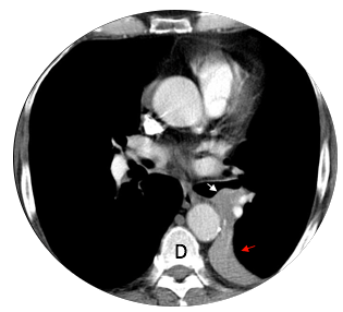

Final diagnosis: carcinoma with LLL collapse
Congratulations to the great majority who made the diagnosis of LLL collapse. Ruqqayia was the first and deserves to be mentioned.
Teaching point: there is little to teach in this case because most of you made the diagnosis. Not everybody is so knowledgeable, though: this patient was seen in the Emergency Room of a large hospital and was told by the attending physician that his chest was unremarkable. Films were rescued by the radiologist the morning after and CT ordered.